 EN

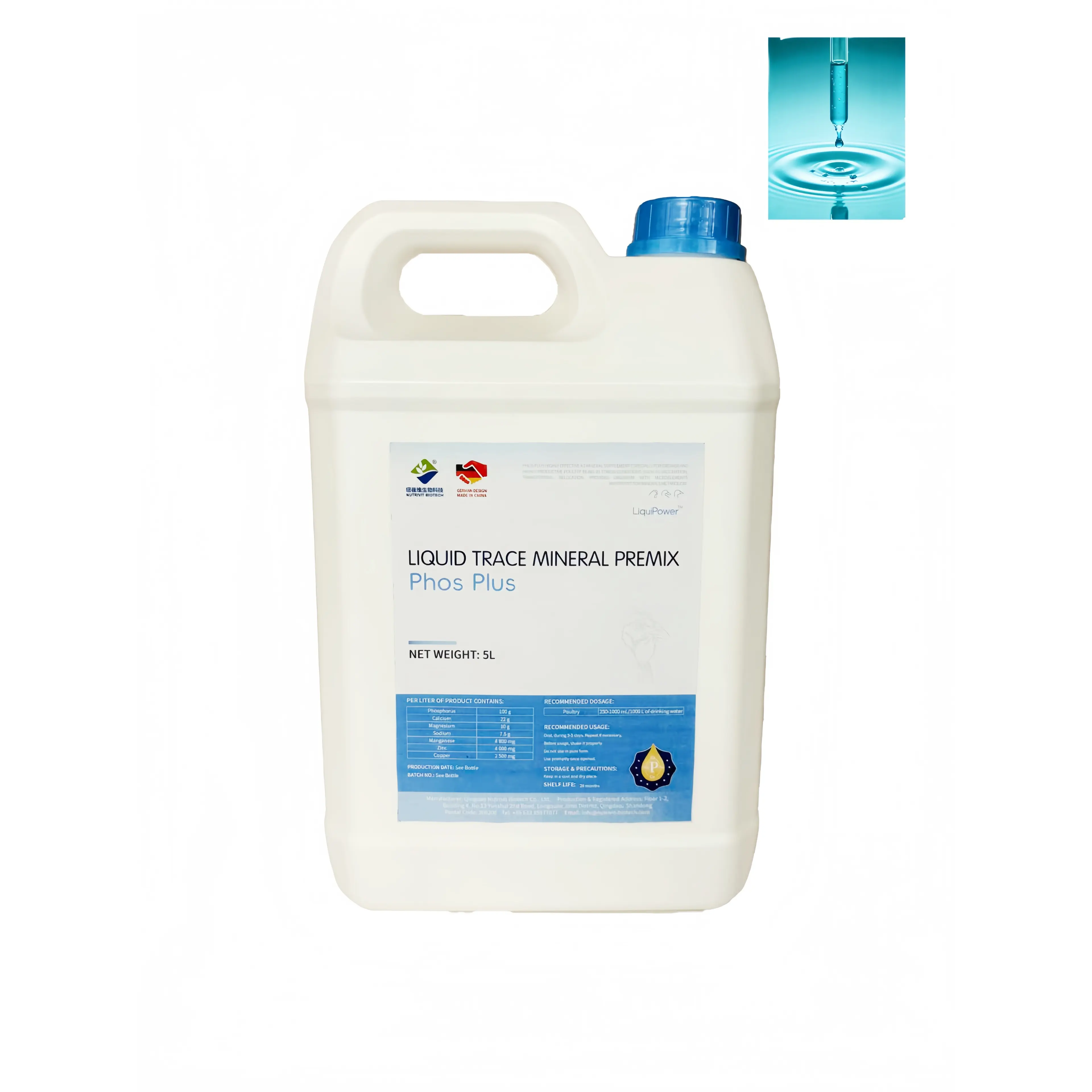
Traces Optimize Intestinal Mineral Absorption and Bioavailability
Trace minerals optimize intestinal mineral absorption and bioavailability by enabling enzyme-driven transport and shielding uptake from dietary inhibitors.
Activation of Metalloenzymes and Transporters (e.g., DMT1, ZIP4) by Copper, Zinc, and Iron
Copper, zinc, and iron cross the intestinal epithelium primarily via metal-specific transporters—not passive diffusion. Divalent metal transporter 1 (DMT1) mediates most duodenal iron uptake, while ZIP4, a zinc-regulated transporter, increases expression during low dietary zinc. Copper-dependent ferroxidases like hephaestin oxidize ferrous iron to ferric iron before its basolateral export, a step essential for systemic iron delivery. Copper also supports metallothionein synthesis—a cysteine-rich protein that buffers intracellular zinc and copper and facilitates their transfer across the basolateral membrane. Critically, these transporters and metalloenzymes are themselves trace-responsive: marginal copper or zinc status suppresses DMT1, ZIP4, and hephaestin activity, trapping iron in enterocytes even when dietary iron is sufficient. Conversely, supplying bioavailable traces enhances transporter expression and improves apparent mineral retention—establishing a self-reinforcing loop where adequate trace status sustains the molecular machinery of absorption. In production animals, this means trace supply functions as a regulator of intestinal absorption capacity, not merely a nutrient input. Controlled feeding studies confirm improved intestinal mineral retention when copper, zinc, and iron are co-supplied in bioavailable forms (2022). Thus, traces act as functional activators of the gut’s mineral transport network.
Impact of Trace Supplementation Form (Organic vs. Inorganic) on Gut Uptake Efficiency
The chemical form of dietary traces directly influences how much mineral reaches systemic circulation. Inorganic sources—such as sulfates and oxides—dissociate rapidly in the gastrointestinal tract and readily bind phytate, calcium, or other antagonists before absorption. Organic trace minerals—chelated or complexed with amino acids or peptides—remain more stable in the gut lumen and can leverage amino acid transport pathways, reducing competition with dietary inhibitors. This stability protects the metal from precipitation and allows lower inclusion rates while maintaining or improving retention. In laying hens and growing livestock, organic forms consistently demonstrate higher apparent absorption than inorganic salts at equivalent trace levels; substituting organic for inorganic sources has maintained performance while lowering mineral excretion. The benefit lies not in delivering more mineral, but in delivering it in a biologically recognized, functionally protected form. Nutritionists therefore assess trace sources by bioavailability—not just total dietary concentration. For growth-focused operations, selecting a stable, chelated trace mineral improves gut uptake efficiency and mitigates deficiency risk during high-demand phases such as rapid skeletal or muscle accretion.
Traces Regulate Hepatic Mineral Storage and Systemic Distribution
The liver serves as the central hub for trace mineral storage, transformation, and release—orchestrating supply according to growth stage, systemic redox status, and endocrine signals.
Selenium and Iron Coordination in Ferritin Synthesis and Redox-Sensitive mRNA Stability
Ferritin synthesis in hepatocytes is governed by the iron-responsive element/iron regulatory protein (IRE/IRP) system. When intracellular iron is abundant, IRP1 acquires an iron-sulfur cluster and dissociates from ferritin mRNA, permitting translation and safe iron sequestration. Under iron-deficient conditions, IRP1 binds the 5′ IRE of ferritin mRNA and blocks translation. Selenium supports this system through selenoproteins—including glutathione peroxidase and thioredoxin reductase—which protect the iron-sulfur cluster of IRP1 from oxidative disassembly. By preserving IRP1 function under redox stress, selenium maintains ferritin mRNA stability and ensures appropriate iron storage capacity. Selenium deficiency disrupts this protection, leading to premature ferritin mRNA decay, reduced hepatic iron storage, and elevated labile iron pools. In growing livestock, adequate selenium status thus enables controlled iron sequestration and timely release into transferrin-bound plasma iron—buffering daily fluctuations in dietary intake, protecting hepatocytes from oxidative injury, and sustaining iron availability for erythropoiesis and muscle development. Selenium and iron do not act independently; their redox-sensitive crosstalk determines both the quantity of iron entering hepatic stores and the kinetics of its mobilization.
Manganese-Dependent Mitochondrial Antioxidant Defense (SOD2) Supporting Liver-Mediated Mineral Release
Manganese is the essential cofactor for manganese superoxide dismutase (SOD2), the primary mitochondrial antioxidant enzyme. Hepatocytes rely heavily on mitochondrial energy for mineral storage, protein synthesis (e.g., ceruloplasmin, transferrin), and ATP-dependent export mechanisms. During these processes, mitochondria generate superoxide radicals; SOD2 converts them to hydrogen peroxide, preventing lipid peroxidation and preserving membrane potential. With sufficient manganese, SOD2 activity remains high enough to sustain mitochondrial integrity and ATP output—enabling efficient copper, iron, and zinc export from the liver. When manganese is limiting, SOD2 activity declines, mitochondria become dysfunctional, and energy-dependent mineral release falters. This results in reduced circulating ceruloplasmin, transferrin-bound iron, and albumin-bound zinc—even if hepatic stores are replete. Therefore, manganese-dependent mitochondrial protection directly couples hepatic mineral reserves to systemic distribution. In fast-growing livestock, maintaining optimal manganese status supports the liver’s ability to release stored minerals precisely when demand surges—for bone mineralization, hemoglobin synthesis, and muscle protein accretion.
Traces Enable Bone and Muscle Mineralization via Endocrine Signaling
Iodine-Driven Thyroid Hormone Activation of Osteocalcin and IGF-1 Pathways
Iodine regulates endocrine signals that coordinate bone and muscle mineralization in growing livestock. The thyroid gland incorporates iodine into thyroxine (T4) and triiodothyronine (T3); T3 binds nuclear receptors in osteoblasts and myocytes to upregulate osteocalcin expression—promoting calcium deposition into bone matrix. Simultaneously, T3 stimulates hepatic and local production of insulin-like growth factor 1 (IGF-1), which drives chondrocyte proliferation, muscle protein synthesis, and satellite cell activation. A 2023 review of mineral metabolism in ruminants notes that this thyroid–IGF-1 axis tightly couples bone formation to lean tissue growth. Marginal iodine intake reduces T3 and IGF-1 output, decreasing calcium retention and increasing urinary calcium loss. Balanced iodine supply restores this signaling loop, improving calcium and phosphorus retention in bone rather than excretion. In beef calves, adequate iodine supports earlier peak bone mass and improved frame development. Trace nutrition programs aligned with species-specific iodine requirements stabilize thyroid hormone output and amplify IGF-1–mediated muscle accretion without over-supplementation—making this endocrine support indispensable for efficient, sustainable gain.
Practical Trace Nutrition Strategies for Growth-Oriented Livestock Operations
Practical trace use must be dynamic—not static. Precision feeding shifts mineral delivery from group averages to individual requirements, and a 2019 review of precision livestock feeding found real-time matching of trace supply to intake, genotype, and production stage reduces feed cost and nutrient excretion.
| Strategy | Key action | Growth-related benefit |
|---|---|---|
| Precision trace supply | Adjust copper, zinc, manganese, selenium, and iodine levels to production stage, intake, and genotype | Supports metalloenzymes, thyroid function, and bone-muscle signaling without oversupply |
| Organic trace forms in high-stress windows | Use chelated or proteinated minerals during weaning, transport, heat stress, or disease challenge | Improves gut uptake and reduces mineral antagonism |
| Feed and water monitoring | Test total dissolved minerals, pH, and antagonists before mixing rations | Maintains target bioavailability and prevents hidden deficiencies |
| Biomarker-based correction | Track liver or serum selenium, zinc, and copper, along with thyroid indicators | Enables early correction before measurable growth loss |
Because trace minerals interact, avoid blanket high-dose strategies. Excess zinc depresses copper status; high dietary iron impairs manganese absorption. Instead, align trace supplementation with actual intake, antagonist load, and growth-phase targets. This approach strengthens the return on trace mineral investment while supporting consistent skeletal development, muscle accretion, and feed efficiency.
FAQ: Traces and Livestock Nutrition
What are trace minerals, and why are they important in livestock nutrition?
Trace minerals such as copper, zinc, selenium, and manganese are essential nutrients required in small amounts. They support critical bodily functions, including enzyme activation, antioxidant defense, and bone and muscle mineralization.
How do trace minerals impact gut absorption of nutrients?
Trace minerals optimize nutrient absorption by activating metal-specific transporters and reducing interference from dietary inhibitors. Organic trace forms can improve bioavailability due to their stability in the gut lumen.
Is there a difference between organic and inorganic trace minerals?
Yes, organic trace minerals are more stable and bioavailable compared to inorganic forms like sulfates or oxides. Organic minerals reduce competition with dietary antagonists, leading to better uptake and retention.
How do trace minerals influence liver function in livestock?
The liver stores, transforms, and releases trace minerals based on growth needs and systemic signals. Adequate trace supply supports efficient mineral release and prevents oxidative stress in hepatocytes.
What are practical strategies for optimizing trace mineral supplementation?
Effective strategies include precision trace supply based on individual needs, using organic trace forms during stress periods, monitoring feed and water quality, and biomarker-based corrections for early deficiency signs.
Table of Contents
- Traces Optimize Intestinal Mineral Absorption and Bioavailability
- Traces Regulate Hepatic Mineral Storage and Systemic Distribution
- Traces Enable Bone and Muscle Mineralization via Endocrine Signaling
- Practical Trace Nutrition Strategies for Growth-Oriented Livestock Operations
-
FAQ: Traces and Livestock Nutrition
- What are trace minerals, and why are they important in livestock nutrition?
- How do trace minerals impact gut absorption of nutrients?
- Is there a difference between organic and inorganic trace minerals?
- How do trace minerals influence liver function in livestock?
- What are practical strategies for optimizing trace mineral supplementation?