 EN
EN

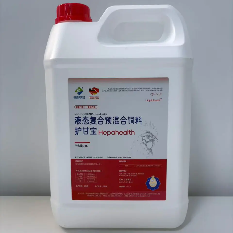
The Liver as the Metabolic Command Center in Livestock
How Liver Metabolism Integrates Energy Partitioning, Detoxification, and Immune Crosstalk
The liver serves as the central metabolic hub in livestock—orchestrating energy partitioning, detoxification, and immune regulation in real time. During high-demand phases like lactation, it directs nutrients toward milk synthesis, growth, or storage based on hormonal cues. A key function is gluconeogenesis: synthesizing glucose from non-carbohydrate precursors (e.g., propionate, amino acids, glycerol) to sustain blood glucose levels when dietary intake is insufficient. Concurrently, the liver manages lipid flux by esterifying non-esterified fatty acids (NEFA) into triglycerides and packaging them into very-low-density lipoproteins (VLDL) for export—preventing intrahepatic fat accumulation. Beyond metabolism, the liver filters portal blood to neutralize endotoxins, ammonia, and xenobiotics, while producing acute-phase proteins (e.g., haptoglobin, serum amyloid A) and cytokine regulators that shape systemic immunity. This tight coupling means hepatic metabolic efficiency directly influences immune competence: a well-functioning liver supports both productive performance and pathogen defense without trade-offs.
Consequences of Dysregulated Liver Metabolism: Fatty Liver, Ketosis, and Immunosuppression in Transition Cows
The transition period—from three weeks before to three weeks after calving—represents the greatest metabolic challenge in the dairy cow’s life cycle. As energy demand surges to support colostrum and early milk synthesis, adipose tissue mobilization floods the liver with NEFA. When VLDL export capacity is overwhelmed, triglycerides accumulate in hepatocytes, triggering hepatic lipidosis (fatty liver). This impairs gluconeogenesis, deepening the energy deficit and driving excessive ketogenesis—manifesting as subclinical or clinical ketosis. An observational study found over 50% of transition cows develop subclinical ketosis, which correlates strongly with suppressed immune function and increased incidence of metritis, mastitis, and retained placenta. The underlying mechanism is multifaceted: lipid-laden hepatocytes show reduced synthesis of complement factors and acute-phase proteins, diminished Kupffer cell phagocytosis, and altered cytokine signaling. Thus, hepatic metabolic failure initiates a self-reinforcing cascade—compromising both productivity and resilience.
Biomarkers and Early Detection of Suboptimal Liver Metabolism
Key Clinical Indicators: Serum NEFA, AST, GLDH, and BHB as Predictors of Hepatic Stress
Blood biomarkers offer a practical, objective lens into peripartal liver metabolism. Non-esterified fatty acids (NEFA) reflect adipose mobilization intensity; prepartum concentrations >0.4 mEq/L indicate excessive lipolysis and predict a 2- to 4-fold increase in postpartum disease risk. Beta-hydroxybutyrate (BHB), measured in whole blood or serum, signals incomplete fatty acid oxidation—≥1.2 mmol/L during the first week post-calving is the accepted threshold for subclinical ketosis. Hepatocellular enzyme elevations add specificity: aspartate aminotransferase (AST) rises with generalized hepatocyte membrane damage (values >100 U/L suggest substantial injury), while glutamate dehydrogenase (GLDH) is highly liver-specific in ruminants and more sensitive to mitochondrial stress—often rising before AST. Together, these four markers map the functional progression: NEFA marks the onset of energy deficit; BHB reveals metabolic inflexibility in fat utilization; GLDH flags early organelle-level compromise; and AST confirms structural hepatocyte damage. Used in combination, they enable detection of hepatic stress well before irreversible histopathological changes occur.
Interpreting Biomarker Trends: Why Timing Matters in Peripartal Liver Metabolism Monitoring
Isolated biomarker values have limited diagnostic power—the trajectory across standardized timepoints reveals whether liver metabolism is adapting appropriately or decompensating. NEFA typically peaks at calving after a steady rise beginning ~10 days prepartum, then declines rapidly as dry matter intake rebounds. Persistent elevation beyond day 3 postpartum indicates failure to re-establish energy balance. BHB follows with a 1–3 day lag; a secondary peak after day 7 suggests impaired ketone clearance or ongoing gluconeogenic insufficiency. GLDH often begins rising subtly during the close-up period—even before NEFA exceeds thresholds—highlighting early mitochondrial vulnerability. Sampling at consistent intervals (e.g., days −14, −7, 0, +3, +7, and +14 relative to calving) allows clinicians to distinguish normal homeorhetic adaptation from pathological drift. For example, a steep GLDH rise between days −7 and 0, coupled with delayed NEFA decline, signals high risk for fatty liver—enabling timely nutritional or management intervention before clinical disease emerges.
Nutritional Strategies to Optimize Liver Metabolism in High-Performance Dairy Cows
Choline and Betaine: Enhancing VLDL Export to Reduce Hepatic Triglyceride Accumulation
Rumen-protected choline and betaine are evidence-based methyl-donor supplements that directly support hepatic lipid export during the periparturient period. Choline is essential for phosphatidylcholine synthesis—the primary phospholipid component of VLDL particles. When methyl donors are limiting, VLDL assembly falters, trapping triglycerides in hepatocytes. Supplementation with rumen-protected choline has been shown to reduce hepatic triglyceride concentration by 30–40% in transition cows (Grummer, 2008). Betaine acts synergistically by sparing methionine and donating methyl groups for the same pathway, enhancing the efficiency of fat packaging and secretion. Field trials demonstrate that combined supplementation lowers the incidence of subclinical ketosis by up to 35% and improves early-lactation milk yield—confirming that supporting VLDL export alleviates a critical bottleneck in liver metabolism.
Methionine and N‑Acetylcysteine: Supporting Glutathione Synthesis and Oxidative Resilience
Methionine and N-acetylcysteine (NAC) target hepatic redox balance—a cornerstone of metabolic resilience during the transition period. Methionine provides sulfur and methyl groups required for glutathione (GSH) synthesis, while NAC delivers bioavailable cysteine, bypassing the rate-limiting step in GSH production. During early lactation, heightened fatty acid oxidation and inflammatory signaling generate reactive oxygen species (ROS); without adequate antioxidant buffering, ROS damage mitochondria, impair gluconeogenic enzymes, and trigger apoptosis. Research shows methionine supplementation increases hepatic glutathione concentrations by up to 25% (Drackley, 2026), and co-supplementation with NAC further amplifies this effect. The result is improved mitochondrial integrity, sustained NEFA oxidation capacity, and lower expression of oxidative stress markers (e.g., 4-HNE, protein carbonyls) in liver tissue. This dual-nutrient strategy strengthens the liver’s ability to manage metabolic load without succumbing to oxidative fatigue—supporting both metabolic stability and immune readiness.
FAQ
What are the primary functions of the liver in livestock?
The liver in livestock plays key roles in energy partitioning, detoxification, lipid metabolism, and immune regulation to ensure optimal productivity and health.
What causes fatty liver and ketosis in transition cows?
Fatty liver and ketosis are caused by overwhelmed VLDL export due to excessive NEFA mobilization during high-energy demand periods like early lactation, impairing gluconeogenesis and leading to lipid accumulation and ketone overproduction.
Which biomarkers are indicators of liver metabolic stress?
Key biomarkers include NEFA, BHB, AST, and GLDH. These reflect energy deficits, incomplete fatty acid oxidation, hepatocyte damage, and mitochondrial stress, respectively.
How can nutritional strategies help optimize liver metabolism?
Supplements like rumen-protected choline, betaine, methionine, and N-acetylcysteine enhance hepatic lipid export, glutathione synthesis, and oxidative stress resilience, improving liver function during the periparturient period.